Medial and Lateral collateral Ligament Injuries
The knee is the largest and one of the most complex joints in the human body. On a yearly basis, near 12 million people visit orthopedic surgeons due to knee ligament injuries. The knee complex consists of 4 bones, multiple ligaments, muscles and tendons, cartilage and soft tissue. Knee ligaments connect bones to bones allowing for stability.
Ligaments
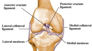
Ligaments of the knee are classified as cruciate and collateral ligaments. Ligaments connect bone to bone. The major ligaments of the knee consist of the ACL (anterior cruciate ligament, PCL (posterior cruciate ligament), MCL (medial collateral ligament) and LCL (lateral collateral ligament). Cruciate ligaments are within the knee joint. They prevent anterior (forward) and posterior (backward) translation of the femur from the tibia. Collateral ligaments are outside the knee joint and prevent medial (inside) and lateral (outside) instability.
Injuries to the collateral ligaments occur with the following stresses – Varus stress to the LCL (inside to outside force) or valgus stress the MCL (outside to inside force). High-risk sports include rugby, football, gymnastics, mixed martial arts and soccer. Many times these injuries in athletics occur from contact. Falls and twisting injuries can also cause damage to these ligaments. Injuries to these ligaments are called tears or sprains. Ligament sprains are classified on a severity scale called grades.
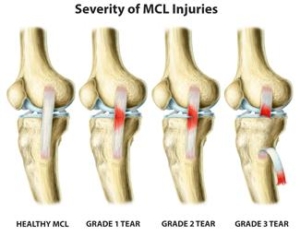
- Grade 1 sprains occur with mild damage to the structure. These injuries continue to keep the joint stable but the ligament is stretched / mildly torn.
- Grade 2 sprains or moderate tears when they occur cause added instability and looseness of the joint. Partial tears at this level may cause episodes or feelings of instability. This grade of injury can be treated without surgery but if typical conservative measures fail then surgery would be recommended.
- Grade 3 sprains or full/complete tears allow for complete joint laxity or no stability typically require surgery to remedy.
Signs and Symptoms
Signs and symptoms of injury to this major ligament can occur in a variety of ways including sports, motor vehicle accidents and falls to name a few. Typically the ligament tears with a sudden and rapid change of direction or stopping. It can also occur with direct impact or a compressive force such as landing. Major symptoms within 24hours can include pain, swelling, lost range of motion and instability with walking or attempted return to activity. Some can even feel a tear or pop in the knee when injury occurs.
Diagnosis
Diagnosis will be determined through a thorough history and physical examination by a trained sports medicine and orthopedic knee specialist. Examination will consist of palpating the knee complex for pain and a visual examination for deformities. The clinician will check both active and passive range of motion. Specific manual tests will be performed by the specialist to determine if inflammation exists, the amount of weakness, instability and other possible factors causing symptoms. Once the initial diagnosis has been made, radiological examinations may need to take place for further assessment. An MRI (3 dimensional picture) will be ordered to assist in determining the severity of the ligamentous injury. X-rays of the knee complex may be ordered to determine if any bony conditions exist such as fracture or loose bodies prior to the MRI.

Normal MCL
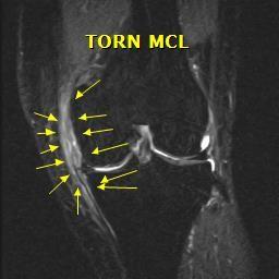
Torn MCL
Treatment
Treatment rarely requires surgery with an isolated MCL sprain and can be resolved through non-surgical techniques. However, multi-ligamentous injuries to the knee may require surgical management.
- Non-surgical management may be indicated and recommend in cases of isolated tears with no additional derangement for patients that have maintained overall stability of the knee following injury. Conservative management of tears can consist of modification of activity and rest, anti-inflammatory medications, a cortisone injection, orthobiologics such as PRP (platelet rich plasma) or Stem Cell injections, bracing and formal physical therapy.
- Surgical treatment requires either reconstruction or repair of the ligament. Surgical repair is performed on high-grade tears or acute full thickness tears. This involves reattaching the MCL or LCL ligament to its insertion site with sutures and surgical anchors. Surgical reconstruction requires your surgeon to replace the torn ligament with a tissue graft. These grafts are usually allografts (donated human cadaver tissue). Graft tissue choices will be discussed with you during your office examination.
Click link for MCL Repair Video
Click Link for LCL Repair Video
Rehabilitation
Rehabilitation is recommended for both surgical and non-surgical management. Regardless of your treatment choice, your sports medicine physician will determine a personalized treatment program that fits your needs. Your active involvement will improve your recovery time. Following the initial onset of signs and symptoms, the initial goal is to reduce swelling and pain, increase mobility and range of motion. At home and at physical therapy, the use of ice, modalities and specific range of motion and strengthening exercises will be performed several times throughout the day. Pain and anti-inflammatory medication may also be prescribed for additional benefit. Crutches and bracing may be initialized following your assessment to aid in stability and prevent further damage to the knee complex. Non-surgical recovery can range from 2-6 weeks for Grade 1-2 MCL and LCL sprains. Non-surgical grade 3 MCL and LCL sprains 6-12 weeks to recover. Once surgical management is completed, physical therapy will begin the following day, working towards restoring full range of motion, strength, and stability of the knee complex. Advancement to a work specific or sports specific program typically occurs at approximately 3-4 months, with full return to sports activities between 4-6 months.