What is Patellofemoral Ligament Reconstruction, Patellofemoral Instability, Lateral Retinacular Release?
Lateral Release
Surgery performed by Dr. Chams
The knee is the largest and one of the most complex joints in the human body. It consists of 4 bones, multiple ligaments, muscles and tendons, cartilage and soft tissue. The boney structures of the knee consist of the patella (knee cap), Femur (thigh bone), Tibia and Fibula (lower leg bones). The patella glides with knee flexion and extension within the trochlear groove. Gliding of this area is accomplished through articular cartilage, one of the two types of cartilage within the knee complex. Articular cartilage is the protective covering over the ends of the bones. This allows the joint to glide permitting fluid motion during active and passive activities.
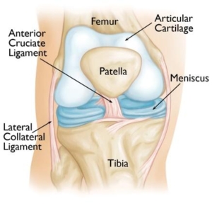
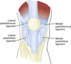
The gliding motion of the patella is stabilized with the additional soft tissue structures such as ligaments, muscles, tendons, synovial tissue and the fat pad beneath the patella. Patellofemoral ligaments are connective bands that stabilize the joint from unwanted movements such as subluxations and dislocations. The lateral and medial patellofemoral ligaments, as seen below, are the main two stabilizing ligaments of the patella. The joint and its components function together to provide stability and power for activities of daily living and athletic movements.
Signs and Symptoms
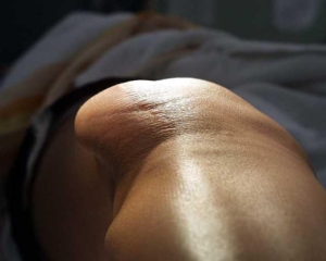
Signs and symptoms of patellofemoral instability are located within the front or anterior aspect of the knee including pain along the patella (knee cap). These injuries can be exacerbated by a traumatic event such as a subluxation or dislocation. Symptoms can also be a result of a genetic disposition to ligamentous laxity or a shallow or uneven trochlear groove. This results in significant pain, swelling and feelings of instability around the patella. Lost motion and the inability to bear full weight is not uncommon. Characteristically, muscular imbalances, muscular tightness or weakness can be a contributing factor to the cause of this injury and condition. When traumatic, the patella either dislocates (comes out of the socket and is reduced by medical staff or persons) or subluxates (moves out of the socket and into socket on its own). Patellofemoral ligaments are either stretched or torn and articular cartilage can be damaged. This can transpire from a direct blow, fall onto the knee or forceful twisting. Once this has occurred there is an 80% to 90% recurrence rate.
Diagnosis
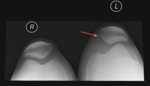
Diagnosis will be determined through a thorough history and physical examination by a trained sports medicine and orthopedic knee specialist. Examination will consist of palpating the knee complex for pain and a visual examination for deformities. The clinician will check both active and passive range of motion. Specific manual tests will be performed by the specialist to determine if inflammation exists, the amount of weakness, instability and other possible factors causing symptoms. Once the initial diagnosis has been made, radiological examinations may need to take place for further assessment. An MRI (3 dimensional picture) may be ordered to assist in determining the injury. X-rays of the knee complex may be ordered to determine the alignment of the patella and depth of the trochlear groove (shown below) or if any additional bony conditions exist such as fracture or loose bodies prior to the MRI.
Treatment
Treatment for Patellofemoral instability will be determined following the initial assessment. Non-surgical and surgical management is based on age, history, anatomy, athletic expectation and extent of the traumatic injury.
- Non-surgical management includes rest, ice, compression, bracing, anti-inflammatory or narcotics medications, decrease or change of activity level until asymptomatic and physical therapy.
- Surgical management through arthroscopy occurs when a small camera is placed into the knee joint to visualize the damage. During this visualization, ligaments, articular cartilage, meniscus and other soft tissue structures are evaluated. Once the assessment has been completed the surgeon uses a variety of instrumentation to repair, or debride (clean) the knee complex. Several procedures can be performed for this condition and are explained below:
- A lateral release is performed to correct the patellar alignment within the trochlea. This occurs when the tissue, lateral retinaculum, is ablated within the knee capsule. The goal following this procedure is to place the patella within its centralized location with a normal tilt within the trochlear groove.
- Medial Patellofemoral Ligament Reconstruction or MPFL reconstruction is an open, minimally invasive procedure performed using an allograft (human cadaver hamstring tendon) or autograft (your own hamstring tendon) to reconstruct the torn ligament injured during the patella dislocation or multiple subluxations. This procedure has a high success rate for chronic patellar instability.
Rehabilitation
Rehabilitation is recommended for both surgical and non-surgical management. Regardless of your treatment choice, your sports medicine physician will determine a personalized treatment program that fits your needs. Following the initial onset of signs and symptoms, the initial goal is to reduce pain, increase mobility and range of motion. At home and physical therapy, the use of ice, modalities and specific range of motion and strengthening exercises will be performed several times throughout the day. Pain and anti-inflammatory medication may also be prescribed for additional benefit. Crutches and bracing may be initialized following your assessment to aid in stability and prevent further damage to the knee complex. Total recovery varies for the condition and the patient. Typically, many will see improvement in symptoms within the first 4-6 weeks following the beginning of treatments. Return to sport or work activity from 6-8 weeks post injury. If surgical management takes place, physical therapy will begin the following day after surgery. A physical therapist and or athletic trainer will work from a specific protocol advancing range of motion, strength, and stability of the knee complex. Recovery of from an MPFL, AMZ – Fulkerson or osteotomy is typically 4 months.